Macular Hole
A macular hole is the development of a small opening in the central part of the macula responsible for central vision. Symptoms range from mild central blurring in the early stages to legal blindness if left untreated.
Macular holes are most common in people over the age of 60 and more often occur in women. The most common mechanism for development of a macular hole is the contraction of the vitreous gel pulling abnormally on the retina to create the hole. Other causes include ocular trauma, diabetic eye disease, myopia, macular pucker (scar tissue contraction over the macula), uveitis, and retinal detachment.
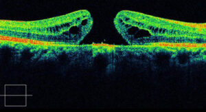
By Yusuke Oshima, from Retina Image Bank, 2013.
Optical coherence tomography or OCT is the preferred method to visualize a macular hole.
Treatment
Vitrectomy surgery is the mainstay of treatment for a macular hole. Small gauge instrumentation is used to remove the vitreous gel that is pulling on the macula. A gas bubble is then injected to bring the edges of the hole together until it heals. Face-down positioning is needed for several days after the surgery depending on the characteristics of the hole. The bubble is gradually replaced by the natural fluid made by the eye. The surgery carries over 90% success rate of hole closure with most patients experiencing improvement in vision.